
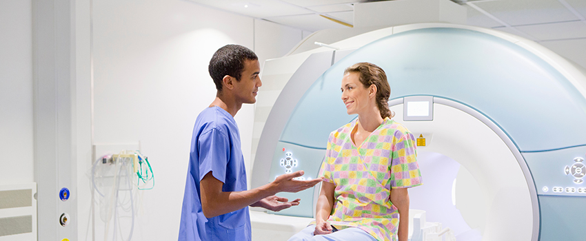
Magnetic resonance imaging (MRI)
We use 1.5 T MRI technology- the standard for producing high-quality images.
What is an MRI?
An MRI (Magnetic resonance imaging) is a test that uses powerful magnets, radio waves, and computer technology to produce detailed pictures of the inside of the body.
Your doctor can use this test to diagnose certain conditions or to see how well you've responded to treatment.
Types of MRIs
At Proscan, we specialise in the following scans:
- Abdomen
- Brain
- Breast
- MSK (Musculoskeletal)
- Prostate
- Spine
- Pelvis
- Joints
When do I need an MRI?
Your doctor may recommend an MRI scan for a number of reasons, including:
- Muscles and Joints - An MRI scan is often used to look at tissue around bones and joints, to help diagnose injuries and conditions of the hip, elbow, and knee. It can also detect conditions such as arthritis, crepitus, bursitis, or tendon tears.
- A heart MRI can capture images of your heart, including the blood vessels and valves, helping to diagnose heart disease or defects. They may also be used to examine the heart following a heart attack.
- A brain MRI can analyse the brain for tumours, abnormal tissues growth and possible causes of a headache. MRIs can also assess any brain damage following a stroke.
- MRIs to detect tumours: MRIs can produce detailed images of soft tissue- these show the difference between normal and diseased tissue. MRIs can also check the progress of a tumour to determine whether it is growing or shrinking.
How to prepare for an MRI
Before an MRI exam, you can eat normally and continue to take your usual medications unless otherwise instructed. We will typically ask you to change into a gown and to remove things that might affect the magnetic imaging, such as jewellery, hairpins, eyeglasses, watches, wigs, dentures, hearing aid, and underwire bras.
What to expect
- The MRI scanner is like a tunnel- similar to that encountered in a conventional CT scanner and is open at both ends.
- You will be asked to lie on your back. Depending on which body part is being examined, you will be placed in the unit, either head or feet first.
- You will be in constant intercom communication with the Radiographer, who also has direct visualisation of you.
- You will hear a knocking sound during the examination- we can supply you with earplugs to reduce the noise.
- In some instances, we need to inject an intravenous injection of a contrast agent. When joints are examined, it may be required for the contrast medium to be injected directly into the joint before the exam.
- You will be given a panic button, which you can use for immediate attention from the Radiographer.
Let our Radiographer know if:
- You are pregnant, or there is any possibility that you could be pregnant.
- You wear a hearing aid, pacemaker, or have an artificial heart valve, cochlear implant, any other implants
- You suffer from claustrophobia.
For best results:
Cooperating with the Radiographer is important for the success of the examination.
You will have to need to remain as still as possible throughout the examination. This will prevent the need to repeat any part of the examination and will also save time and reduce any possible discomfort.